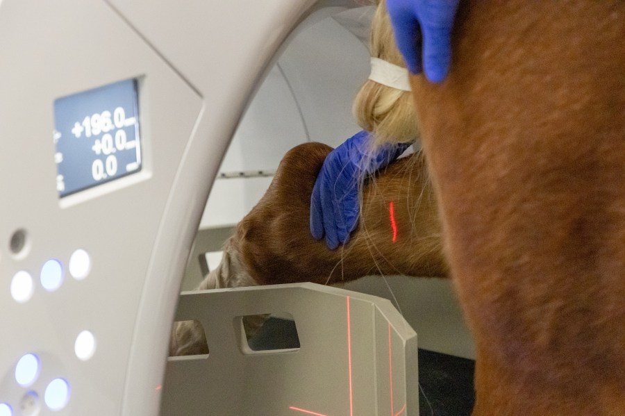
A new advanced Computed Tomography (CT) scanner has been installed in the Royal Veterinary College’s (RVC) Equine Referral Hospital which will allow vets to better diagnose and treat conditions in horses.
The new machine will allow the RVC team to scan parts of a horse’s body that they were previously not able to such as the head and upper part of the neck.
This will help diagnose teeth and sinus problems as well as support the further evaluation of horses who sustain trauma to their head or show signs of headshaking.
The team will also be able to obtain images of the horses limbs up to and including the carpus (knee) and tarsus (hock). This will be useful when they are diagnosing causes of lameness, due to issues with both the bone and soft tissues.
Improved imagery
“This CT is an improvement on what could be done previously,” said Freddie Dash, RVC lecturer in equine diagnostic imaging.
“To begin with, the horse is now standing on the ground and the CT machine is moving. That means the horse is more likely to be still during the examination, resulting in better-quality images.
“The machine also has a larger diameter to its bore, which means that we can fit more of the horse’s anatomy into the machine and scan areas that weren’t previously possible.”
Qalibra Exceed is an advanced scanner which uses a fan beam CT (Canon Aquilion Large Bore Exceed) mounted on a custom platform.
“The welfare of our patients will be massively improved because we can now use the CT to get a more precise and potentially earlier diagnosis,” said Dr Dagmar Berner, senior lecturer equine diagnostic imaging at the RVC.
“For example, to diagnose joint disease and cartilage defects, previously we were able to conduct an MRI or ultrasound in a standing fashion, however, these modalities have their limitations.
“CT is very sensitive for the diagnosis of cartilage defects. If we have a diagnosis earlier on, we can plan a more precise treatment and catch disease early.
“Additionally, our new CT system has significant benefits for students, residents and interns [postgraduate trainees], providing better training and understanding of certain disease processes, as well as the anatomy of the distal limbs.”
Main image credit: Royal Veterinary College


