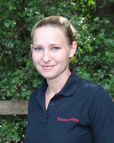
 Kate Southorn from Scarsdale Veterinary Group, a member of XL Vets Equine, tells us about periodontal disease.
Alongside being a general practitioner, I run practice-based dental clinics for patients with more complicated dental disease.
Equine dentistry is one of the most rapidly advancing areas of veterinary medicine. While routine examinations and floating still make-up the majority of the caseload, there are now a range of treatment options including dental bridges and fillings – yes, we are still talking about horses’ teeth!
I see horses at the clinic because they’ve gone off their food, have a nasal discharge or difficulty chewing, but others have subtle signs of serious dental disease identified at routine dental examination.
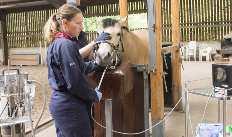
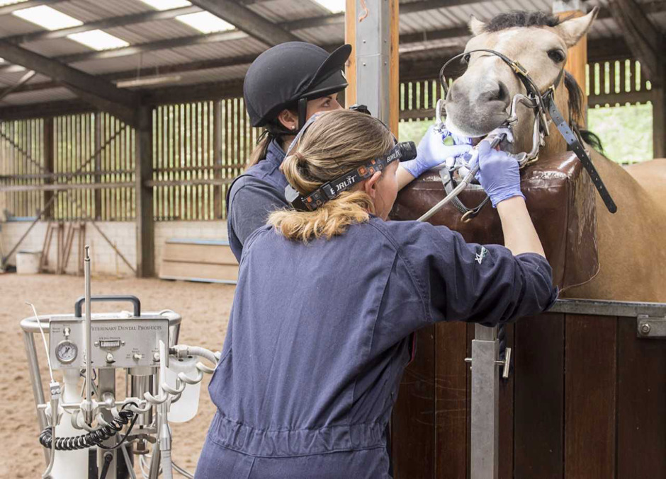 I use sedatives to assist with dental examinations and treatment, this allows the teeth and gums to be examined more carefully, by reducing movement and increased patient relaxation.
A head light and mirror or dental endoscope can then be used to assess the teeth and gums.
One of the most painful conditions I treat is diastema (gaps between the teeth) where food becomes trapped causing periodontal (gum) disease.
 Horses’ cheek teeth are angled to keep a close contact between them, so they function as one unit for grinding food.
When angulation is poor or teeth develop too far apart or are displaced, diastema develop.
In older horses, diastema may develop as the teeth wear away because they become smaller in cross-section, more mobile and may become displaced.
Wide gaps are open and don’t tend to cause problems as food doesn’t get trapped. Narrow gaps act as valves, the food is forced into the gap at the grinding surface and works its way to the gum firmly trapped between the teeth.
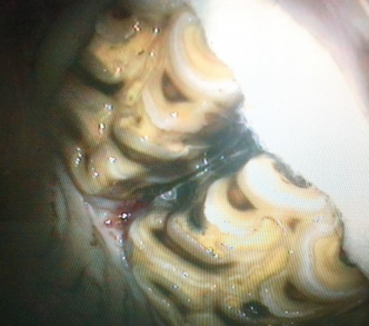
As the food rots, bacteria produce acid that decays the adjacent teeth and damages the periodontal tissues leading to gum recession.
Deep pockets can form if the gum recession is severe enough to affect the underlying bone.
Treatment involves removing trapped food and decayed dental tissue, making sure the teeth are appropriately floated (as the condition is associated with uneven tooth wear).
Temporary packing using dental impression material, acts as a bandage over the damaged gum damage allowing it to heal.
When severe gum disease is present, the gap can be widened to stop food trapping and allow food to be removed from deep between the teeth.
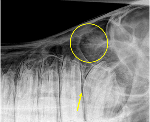
In cases like this one, the food trapped between the upper teeth had worked its way between the teeth (yellow arrow in the X-ray below) into the sinuses and caused a stinking sinusitis. Food was visible on the X-ray image as a mottled white shadow (circled).
I trephined the sinuses, making a small window into the skull over the sinuses, removing the rotting food and a tube was used to flush them with saline twice a day until they were clear of infection.
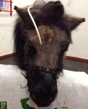
The surgery sites healed within a few days of tube removal. I created a dental bridge to seal the widened gaps using human dental products used to fill teeth.
Now fully recovered, this lovely little chap has returned to his day job visiting residential homes and competing in mounted games and handy pony.
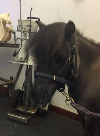 He also helps out at client education days. I regularly examine his mouth, as the teeth continue to erupt and wear away, so will the bridge and it needs to be replaced every two years or so.


