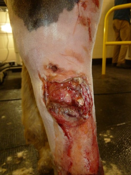
Jack’s wounds on day one
Aoife from XLVets Equine talks us through what happened when a horse called Jack got himself tangled up in some barbed wire.
Horses and barbed wire never mix well. I was reminded of this a few months ago as I stood with a bleeding horse and an upset owner in the middle of a field planning a course of action.
Jack, a skewbald gelding, had been feeling greedy and decided that the grass was greener on the other side of the fence.
Unfortunately, this fence was made of barbed wire and in his eagerness to graze the neighbour’s field, he hadn’t quite jumped high enough and had entangled his right hind leg in the upper strand.
When his owner found him he was standing quietly, looking very sorry for himself in a pool of clotted blood. She immediately called our practice and I arrived half an hour later.
I sedated Jack and did a preliminary examination of the affected leg.
With any wound on the limb of a horse our primary concern is whether or not a synovial structure like a joint or tendon sheath is involved.
As Jack was a typical cob with feathery legs, I had to clip off a lot of hair before I could see exactly what I was dealing with.
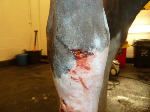
Jack’s wounds on day one
After I’d washed off the dried blood and mud, I found two quite different wounds. One was a few centimetres wide, deep and frankly hideous on his upper cannon.
I would’ve missed the second one if I hadn’t clipped the hair away because it was a small and seemingly innocuous puncture wound.
But this second wound was actually the more sinister of the two. Why? Because it was exactly overlying one compartment of his hock – the tibio tarsal joint to be exact and also worryingly very close to the extensor tendon sheath.
This was an ‘uh oh’ moment for me and the client started crying.
If either of these structures had been entered and contaminated with bacteria, a course of antibiotics wouldn’t be enough – he’d need a general anaesthetic and lavage to literally flush out the infection.
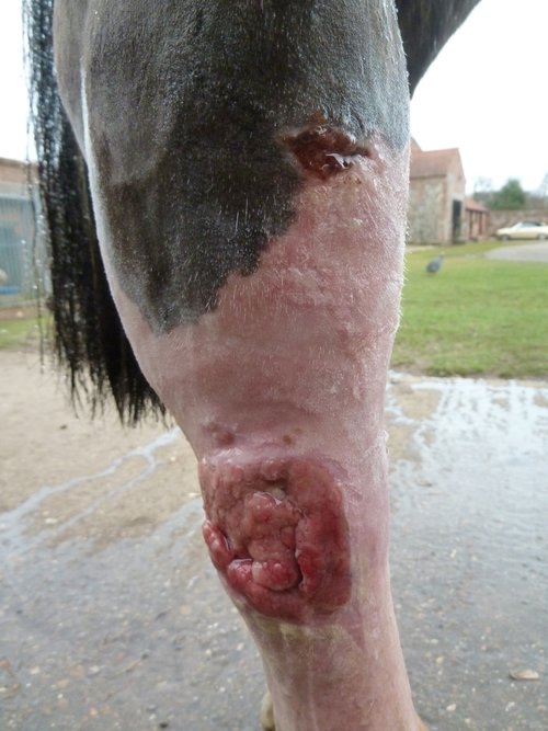
Proud flesh is a common problem in horses
We agreed to admit Jack to our clinic for further investigation. An ultrasound examination of the area followed by a synovial sample (joint tap) revealed Jack was a very lucky horse – the wire had literally missed the synovial structures by a millimetre!
The skin loss over the large wound was too much to bring the edges together for suturing so instead this wound would have to be dressed and bandaged and healed by the process known as second intention.
The wound bed gradually fills up with granulation tissue and slowly but surely healing takes place.
Unfortunately, in horses a common problem is the formation of exuberant granulation tissue otherwise known as ‘proud flesh’.
In Jack’s case this is exactly what happened after a few weeks into his healing process.
Proud flesh inhibits the contraction of the wound so we had to deal with it firmly.
This involved what the owner described as ‘slicing and dicing’ and it was as gruesome as it sounds!
Quite literally the ‘flesh’ is cut away in a surprisingly painless process with a scalpel blade until it no longer sits proud from the wound.
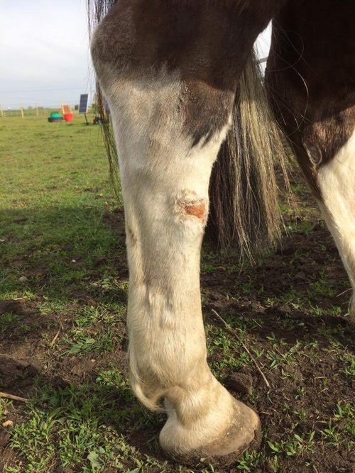
Jack is well on the mend three months later
Every day the owner applied a veterinary-prescribed gel that has a special formulation designed to inhibit the formation of further exuberant granulation tissue and allow the wound to heal slowly but surely.
It has now been three months since Jack’s injury and the wound has almost but not quite healed completely.
Since he’s 100% sound, he gets ridden daily by his owner. She’s certainly learnt her lesson and now ensures he grazes nowhere near any barbed wire fences!


