Splints are bony enlargements (exostoses) of the interosseous ligament that connects the splint bones to the cannon bone in a horse’s leg. They can occur throughout the year, but there is a higher incidence during the summer months.
A splint may be unsightly, but with careful management they don’t usually cause a horse too many problems and won’t lead to a large claim on your horse health insurance policy.
What is a splint?

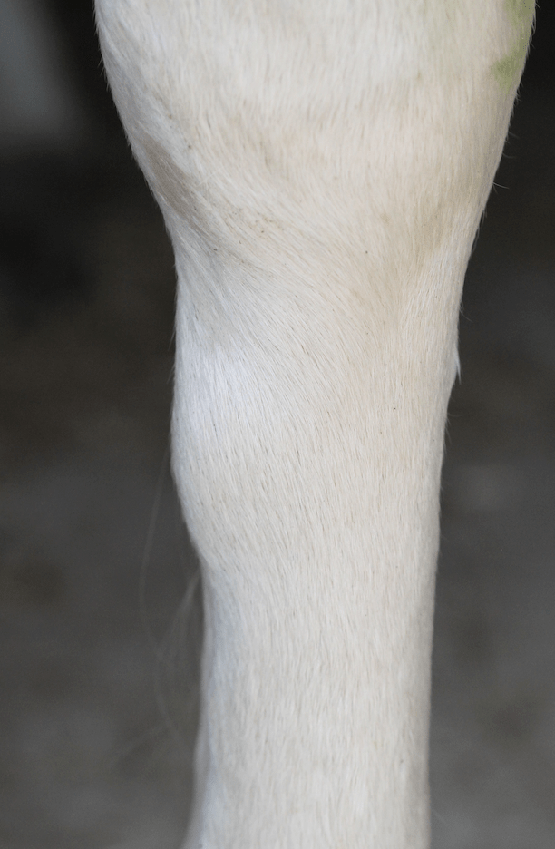
A splint appears as lump on the inside of the leg
Inflammation and mineralisation of the ligament causes a surface bone reaction to the splint and cannon bones, resulting in the development of small to large bony lumps which can be felt around the splint bone region of the lower limb.
For the most part, splints are cosmetic blemishes that don’t interfere with a horse’s long-term athletic ability. However, some can result in significant lameness, especially in the immediate injury period or, in rare cases, where there is impingement of the suspensory ligament.
Each limb has two splint bones: the medial (second metacarpal bone, MC2); and the lateral (fourth metacarpal bone, MC4), which course on either side of the back of the cannon bone.
Splints generally affect the top third of the affected bone, but they are certainly not limited to this area and can occur in any part throughout the length of the bone.
The medial (inside) forelimb splint bone is most commonly affected, but splints may occur on the lateral (outside) forelimb splint bone, or indeed on either hindlimb splint bone, although this is less common.
Diagnosing a splint
Splints can occur as a result of trauma, foot imbalance, or secondary to poor knee conformation, namely bench knee. Young horses are more commonly affected, but splints can affect horses of any age.
Clinical signs vary in severity. Some horses show no evidence of pain or lameness when they have a splint, whereas others do appear lame, have soft tissue inflammation and pain on palpation.
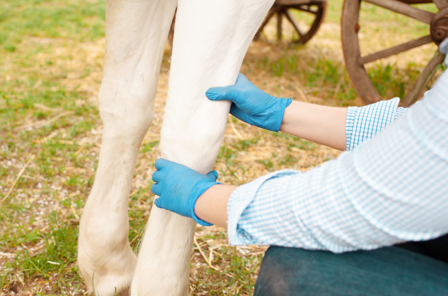
If you notice that your horse has a splint, I recommend putting them on box rest until you have consulted your vet. If your vet examines the horse, they will do so at rest and will carefully palpate the splint to try to discover the likelihood of suspensory ligament impingement.
Your horse will then be trotted up to determine whether they are showing any signs of lameness. If they are lame, nerve blocks may be performed to confirm that the lameness originates from the splint and not from elsewhere.
Managing a splint
A decision will then be made as to whether conservative management is likely to be sufficient, or whether further imaging is required. Further imaging generally involves radiology (X-rays) with or without ultrasound assessment of the suspensory ligament.
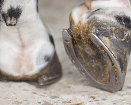
In cases where suspensory ligament involvement seems unlikely, affected horses are generally placed on box rest and given oral anti-inflammatory therapy until the pain and inflammation subside and the lameness clears up. A controlled exercise programme can then begin. Remedial trimming and farriery may be recommended too.
In cases where the suspensory ligament is involved, prolonged box rest will be needed, with your vet evaluating your horse at regular intervals. In those cases that don’t respond to treatment, surgery may be required.
Prognosis for horses with a splint
With time, splints will generally reduce in size, but the timeframe varies from weeks to months, and the new bone formation may never fully resolve. Topical anti-inflammatories may be recommended by your vet to assist with soft tissue inflammation, but timely box rest and oral anti-inflammatories are the most important steps in the management of splints.
Your vet may also recommend hydrotherapy in the initial phase of injury. Distal limb compression, such as the use of a stable bandage and gamgee pad for up to 12 hours daily, may help to reduce the size of the splint.
In the past, other non-orthodox treatments have been used, such as firing and blistering, but these are no longer recommended and nor are they ethical.
Case study: painful to touch and lame

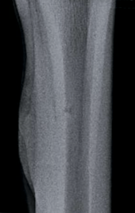
X-ray showing Daisy’s splint (swelling on left side of the bone)
When Daisy, a five-year-old Irish Sport Horse, arrived at Oakhill Equine Vets, her owner had noticed a medial splint on the mare’s left fore 10 days earlier. Palpation of the splint revealed a warm, hard swelling that was painful to touch.
The horse’s suspensory ligament border lacked definition, which raised concerns that there was ligament impingement. She had long-toe/low-heel conformation and, at walk, landed lateral hoof wall first, showing lateromedial imbalance. In trot, I could see mild lameness on the left fore on a straight line and moderate lameness when lunged on a hard surface.
Nerve blocks confirmed that the lameness was caused by the splint. Ultrasound also confirmed that the new bone extended inwardly as well as outwardly, and this resulted in suspensory ligament inflammation (desmitis).
In the first instance, I advised box rest alongside eight weeks of controlled, in-hand exercise. I also prescribed a short course of anti-inflammatories and recommended remedial farrier to improve Daisy’s hoof conformation and balance.
Removing the splint bone
Initially, Daisy improved. However, six weeks into her recovery she suffered a setback which resulted in reoccurrence of swelling and significant lameness. When there was little improvement six weeks later, we repeated the ultrasound and also did an MRI scan. This led to the decision to remove the affected portion of splint bone.
Fortunately, both the surgery and post-operative period went well. Six months later, a small bony nodule was palpable at the surgery site but Daisy was not lame. She began gentle ridden work and three months later returned to eventing.
Images: copyright Shutterstock. Diagram: copyright Leona Bramall/Oakhill Equine Vets


